Ampullary balloon dilation
10 January 2020
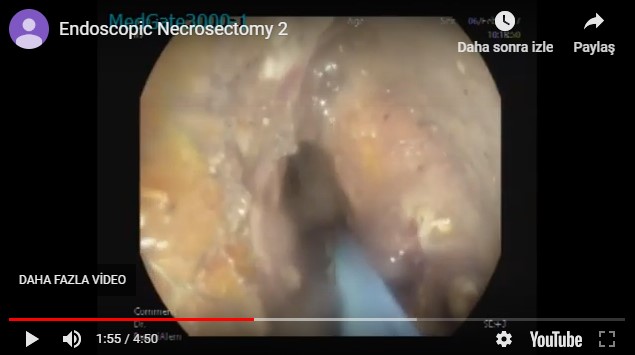
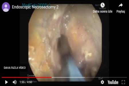
Pancreatic necrosis happens in 20 % of the patients with acute pancreatitis. It may be intrapancretic, extrapancreatic or both. However most of the necrotic collections are sterile and resolves spontaneously. In some patients necrotic tissue is spontaneously infected by the bacterial diapedesis form intestine. Infected necrosis is more common in large necrotic areas. In the past surgical necrosectomy was the standard of care in infected necrosis. Recently endoscopic necrosectomy emerged as an minimal invasive alternative. In these three cases, we demonstrate this procedure.
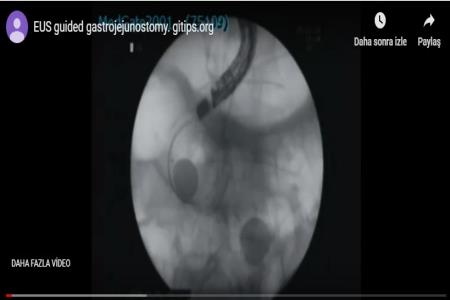
Technique: We approach with a linear therapeutic linear echoendoscope. We puncture the necrotic area with a 19 G needle. We obtain aspirate and send it for microbiologic examination. We insert 0.035 Guide wire, withdraw the needle and dilate the tract with 6 F cystotomy. Afterwards we usually insert a lumen apposing stent of 14-16 mm diameter and lavage the area with hydrogen peroxide which makes furher necrosectomy more effective. It would be better to wait for the full opening of the stent for some days to start necrosectomy. However th clinical status requires immediate necrosectomy may be performed either directly or after baloon dilation of the stent. For necrsoectomy we insert a standard gastroscope into the necrotic cavity. We use CO2 instead of air to prevent air embolism that is very rare complication. We grasp the necrotic debris with biliary stone basket, colon polyp retrieval net and take it out to the stomach and leave there. Sometimes we severe the ncerotic debris with snare with or without administration of electrical current. Slight bleeding always happens. Severe bleeding is rare that is due to the injury of a major artery or vein. If you suspect a vessel in the vicinity of necrotic area, you should avoid from this area. This process may take up to one hour or more. It may be done either under deep sedation or general anesthesia.�
Aa fistula is formed between necrotic area and stomach as early as on week. However development of a fully developed fistula may take 2-3 weeks. In this case we remove the stent, dilate the tract with a baloon (12-18�
 |
10 January 2020
29 April 2019
27 April 2019
24 April 2019
24 April 2019
21 April 2019
3 April 2019
Related posts